A Unified 3D solution for teaching anatomy
TECHNICAL SPECIFICATIONS:
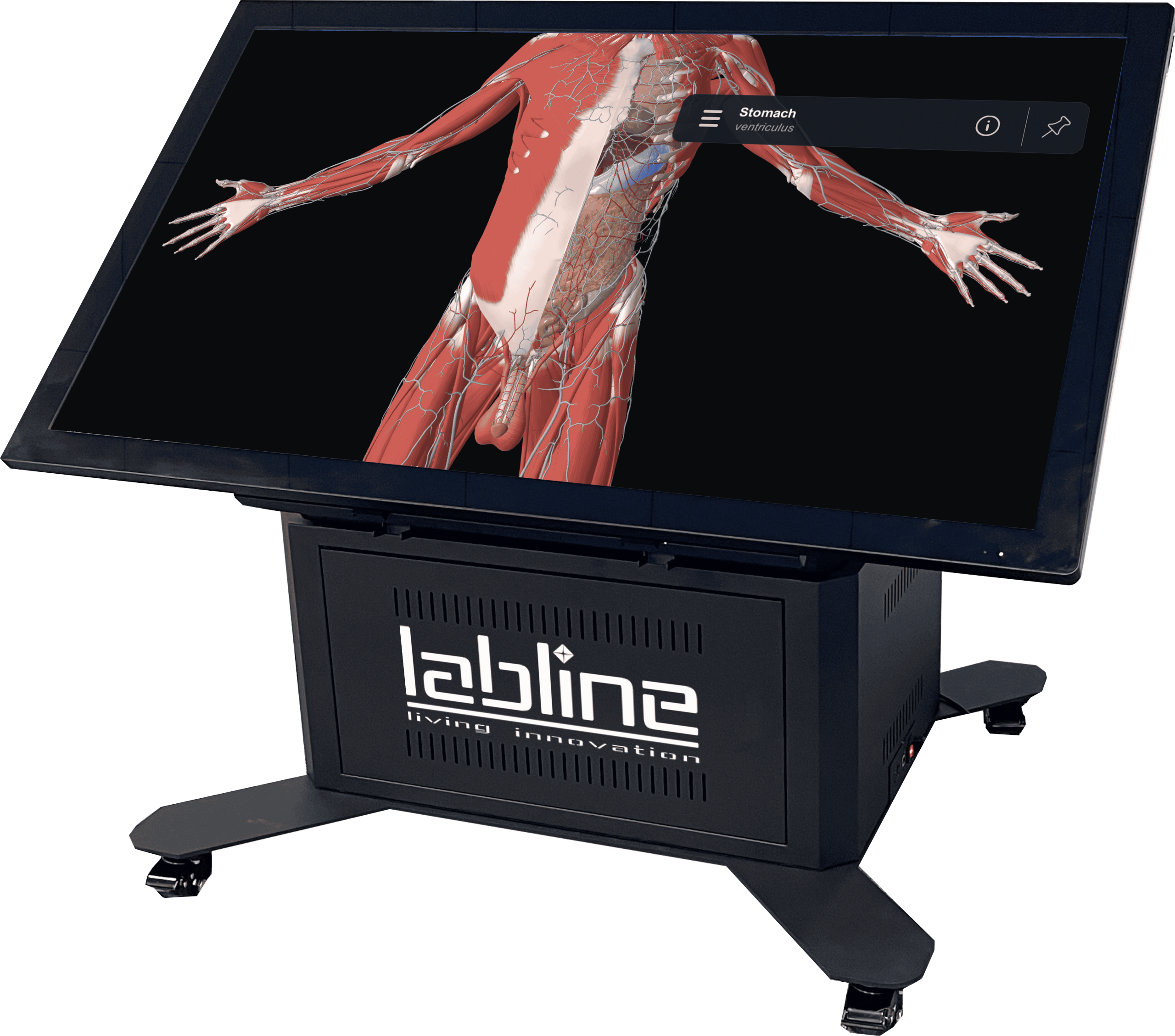
Screen size: 65 inches
Resolution: 4K (3840×2160)
Compatibility: Windows 10 pro & Windows 11 compatible. Compatible with all types/formats of medical images (including pathology images).
Touch points: 20 simultaneous points
Placement/Mobility: Motorized tilting for table position, and motorized elevation for height adjustment – Should be able to be used as Vertical (Lecture Mode) & Horizontal (Dissection Table Mode). Along with heavy duty wheels with locks for easy movement and placement.
Connectivity: Possibility to connect to local network with private and shared spaces. Possibility of connection to projector/external screen.
Female & Male 3D Interactive Model
Regional Anatomy:
Taking a step-by-step approach to studying human anatomy helps you really grasp the complexity of the body’s structures. It deepens your understanding and sharpens your hands-on skills, making it an essential part of learning in the medical field.
Human Anatomy:
Dive into the study of anatomy by focusing on each system one at a time—like the circulatory, respiratory, and nervous systems—to gain a deeper understanding of how they work and connect within the body.
Histology:
You can examine the histology of organs directly on a human model to get a closer look at their microscopic structure.
Pathology & Histopathology:
Teachers can compare healthy organs with diseased ones to better understand pathology. Additionally, the histology of certain organs is accessible for closer examination.
Radiology:
Building a solid understanding of CT scans and MRI scans is essential for interpreting medical imaging. These tools allow us to view detailed cross-sectional images of the body, providing insights into both normal and abnormal anatomy. By learning how to analyze these scans, you can enhance diagnostic accuracy and deepen your knowledge of various medical conditions.
Functional Anatomy:
Discover the wonders of functional anatomy with vivid visuals and clear explanations that showcase how different structures work together to sustain essential functions. It’s a valuable resource for students, professionals, and anyone eager to learn more about the human body.
Digital Dissection/Cadaver Lab:
Our atlas includes a state-of-the-art feature that allows for virtual dissections and provides interactive 3D anatomy models. These tools create an engaging and immersive learning experience, perfect for deepening your understanding of human anatomy.
Scene saving & sharing so you can use anywhere & at any time, Knowledge test, Segmentation, 3D drawing on the model, Preloaded scenes of dentistry & Administrative portal for institutes.
Computer specifications: CPU-Intel i7 12th gen, GPU-RTX 4070TI 12K, Memory-32GB DDR4, Storage 1×256 GB SSD & 1 x1Tb hard disk
Inbuilt and importable training cases: Availability of inbuilt training cases for major medical disciplines including Anatomy, Histology, Radiology, Diagnostic Imaging (Pathology) amongst others. Availability of integrated curriculum of virtual human dissector with option of remote access. Availability of scene saving & sharing among the license holding students and faculty members.
Movement:
4 pieces of heavy-duty wheels, with locks
Software Details:
- Atlas – Life time subscription
- unlimited time software license, lifelong free updates
- Optional Licenses for Students
- Optional Licenses for Teachers
Features:
- Access is readily available during the class without the need for internet connectivity through the use of a Crypto Key
- User Friendly Interface
- Atlas App for Windows, IOS, Android
- Connect to Video Screen and Projectors
- Share Anatomy Scenes with your Students
- Ideal for Distant Learning
- Use Atlas Software to prepare lectures at any time & any Place
- Create Scenes and assess knowledge easily
- 0-90⁰ Tilt & height adjustment
- Smart short-backup UPS to prevent electricity jerk
- 6mm tempered glass
- Handset easily accessible for teacher
- Wireless remote for tilt & lift.


Reviews
There are no reviews yet.