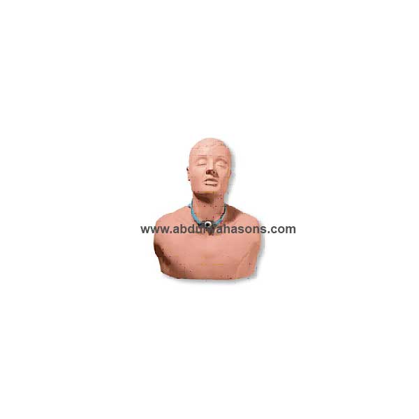
Adult Patient Education Tracheostomy Care Manikin
Product Description
- Adult Manikin
• Dressing Changes
• Cuff Inflation
• SuctioningIdeal for teaching patients and caregivers the skills they will need to perform at home. Includes oral and nasal passages, the pharynx, epiglottis, trachea, esophagus, tracheostomy site, and cricoid cartilage. The mouth and jaw are flexible to allow oral suctioning. The trachea branches into the left and right bronchial trees. Students can practice suctioning techniques, proper cuff inflation, dressing changes, and other techniques. Cleansing and maintenance of the external tracheal site can be performed just as on an actual patient. The adult manikin comes with one tracheostomy tube, instruction guide, and hard carry case.
Related products
-
Code Simulator 12-Lead Arrhythmia Simulator – ZOLL
Pace and defibrillate directly into this interactive 12-lead ECG simulator through your hands-free defibrillation cable. Connects directly to monitor • Does not work with manikin • Simulate electrical capture with your external pacer. Press simulator capture key to select one of four preset pacing capture levels: 70, 80, 90, or 100mA. When pacer current is greater than selected capture level, paced beats appear on your monitor. Waveforms for pacing include: Sinus Brady (two), 1st degree A-V block, 2nd degree type I A-V block, 2nd degree type II A-V block, 2nd degree type II A-V block with PVCs, and 3rd degree A-V block. • Simulate cardioversion with your manual, semi-automatic, or automatic defibrillator. Activate “convert” feature to select - before defib discharge - another rhythm to run immediately after defib discharge. Waveforms for defibrillator training include: V. Fib., V. Tach (high rate), V. Tach (low rate), Torsade, A. Fib, A. Flutter, PSVT, Sinus Tach, Sinus Rhythm, Sinus Rhythm with PVCs, Asystole, and NSR. • Generate realistic 12-lead ECG rhythms. Connect your ECG cable to simulator’s 10 ECG snaps. Independent chest lead and limb lead ECG signals create realistic 12-lead ECGs for each rhythm. Generate ST segment and T wave abnormalities including: Anterior MI, Inferior MI, Antero-Septal ST Elevation, Anterior ST Depression, Lateral ST Elevation, and Inferior ST Elevation. • LED indicators: pacer pulse detection, defib discharge detection, and low battery.
-
Infant Venous Access Simulator
Product Description
A unique, anatomically correct representation of a 4-lb., 16″ female baby developed to teach and practice the vascular accessing of newborns and infants. Practice accessing, securing, dressing, site care, and maintenance for standard venipuncture, central catheters, PICC lines, and umbilical 5 FR catheters.
• Access the median basilic and axillary veins on the right and left arm
• Right leg – saphenous and popliteal veins
• Neck and head – external jugular and temporal (scalp) veins
• Realistic flashback of simulated “blood” confirms proper needle location in the vein
• Redesigned umbilicus allows repeated catheterization of the umbilical catheter, with proper placement verified by a blood return
• Nasal and oral openings allow placement of nasal cannulas, as well as endotracheal, nasogastric, and feeding tubes to teach suctioning, securing, dressing, cleansing, care, and maintenance
Nita Newborn™ includes female baby body; umbilicus; translucent skin on arm, leg, and head; I.D. bracelet; diaper; vein tubing with umbilical valve; blood reservoir bag with attached tubing; blood concentrate; instruction manual; and carry case. -
Advanced Venipuncture and Injection Arm – Light
Product Description
This revolutionary training arm provides complete venous access for IV therapy and phlebotomy, plus sites for intramuscular and intradermal injections. An extensive 8-line vascular system allows students to practice venipuncture at all primary and secondary locations, including starting IVs and introducing Over the Needle IV Catheters. The venous system simplifies setup with only one external fluid bag supplying artificial blood to all veins simultaneously. The dorsal surface of the hand includes injectable metacarpal, digital, and thumb veins. The antecubital fossa includes the median cephalic, median basilic, and median cubital veins. Venipuncture can also be performed along the basilic, cephalic, accessory cephalic, and median antebrachial veins. Intramuscular injections may be performed in the deltoid muscle and intradermal injection sites are located in the upper arm. Intramuscular injections into the deltoid muscle are enhanced by the soft, lifelike skin and by the natural bony landmarks in the region. Intradermal injections using distilled water will create characteristic skin welts at designated sites on the upper arm. The realism of the Life/form® Advanced Venipuncture and Injection Arm is truly amazing. The soft, flexible fingers are molded separately with extreme attention paid to every detail – right down to the fingerprints! Flexion of the wrist helps students develop manipulation skills. The replaceable skin rolls as the veins are palpated, and a discernable “pop” is felt when entering the veins. The Life/form® molding process reproduces the fine details of the skin to make the arm look and feel alive. Valves in the veins can be seen and palpated on the skin surface. Simulated veins and skin are completely replaceable to keep this training arm looking and working like new. Under normal use, hundreds of injections may be performed before the veins or skin need to be replaced. Complete replacement kits are available and easy to use. To extend the life of the veins, an aerosol sealant is available to seal punctures and prevent leakage.
Complete Venous Access:
• Basilic V.
• Cephalic V.
• Digital V.
• Dorsal Metacarpal V.
• Median Basilic V.
• Accessory Cephalic V.
• Extensive Venipuncture System
• Flexible Fingers and Wrist
• Intradermal Injections
• Intramuscular Injections
• Median Antebrachial V.
• Median Cephalic V.
• Median Cubital V.
• Replaceable Skin and Veins -
Code Simulator 12-Lead Arrhythmia Simulator – Phillips
Product Description
Pace and defibrillate directly into this interactive 12-lead ECG simulator through your hands-free defibrillation cable.
• Simulate electrical capture with your external pacer. Press simulator capture key to select one of four pre-set pacing capture levels: 70, 80, 90, or 100mA. When pacer current is greater than selected capture level, paced beats appear on your monitor. Waveforms for pacing include: Sinus Brady (two), 1st degree A-V block, 2nd degree type I A-V block, 2nd degree type II A-V block, 2nd degree type II A-V block with PVCs, and 3rd degree A-V block.
• Simulate cardioversion with your manual, semi-automatic, or automatic defibrillator. Activate “convert” feature to select – before defib discharge – another rhythm to run immediately after defib discharge. Waveforms for defibrillator training include: V. Fib., V. Tach (high rate), V. Tach (low rate), Torsade, A. Fib, A. Flutter, PSVT, Sinus Tach, Sinus Rhythm, Sinus Rhythm with PVCs, Asystole, and NSR.
• Generate realistic 12-lead ECG rhythms. Connect your ECG cable to simulator’s 10 ECG snaps. Independent chest lead and limb lead ECG signals create realistic 12-lead ECGs for each rhythm. Generate ST segment and T wave abnormalities including: Anterior MI, Inferior MI, Antero-Septal ST Elevation, Anterior ST Depression, Lateral ST Elevation, and Inferior ST Elevation.
• LED indicators: pacer pulse detection, defib discharge detection, and low battery.

Reviews
There are no reviews yet.