Advanced Childbirth Simulator
Product Description
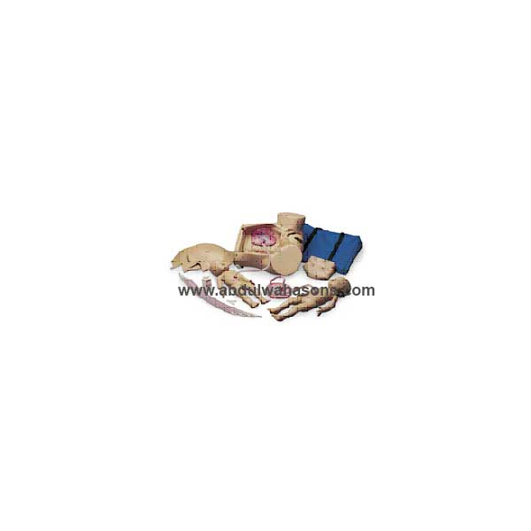
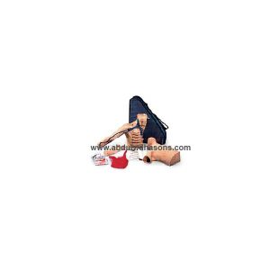
A total simulator for demonstration of all standard obstetrical procedures:
• Fetal Palpation
• Breech Birth
• Normal Birth
• C-Section Delivery
• Episiotomy
• Vertex Presentation
• Prolapse of Umbilical cord
Features include a removable diaphragm end plate and removable stomach cover for manual positioning of fetus; life-size pelvic cavity with all major anatomical landmarks; ultra-soft vulval insert for episiotomy exercises; simulated dorsosacral position for realistic delivery technique. Cannot do the Leopold procedure on this simulator. Forceps can be used during simulation. All parts in this simulator are replaceable. The Advanced Childbirth Simulator with vinyl skin includes two fetal babies with umbilical cords and placentas, spare stomach cover, two vulva inserts, four spare umbilical cords, two umbilical clamps, talcum powder, instruction manual, and soft carrying bag.
Related products
-
SMART STAT Basic with iPad
Product Description
This affordable patient simulator takes ALS training into the students’ environment. Instructors can evaluate student knowledge, skill levels, and critical thinking abilities by utilizing the applications via the iPad®*. This simulator can function in the lab and in the field for ascertaining diagnostic ability. Student performance records can be transferred to a computer, and chronological scenario event logs can be printed. SMART STAT Basic has an on-board air compressor, includes a free app on the iTunes Store®* (SMART STAT), storage for students’ performances, spontaneous breathing, pulses, blood pressure, carotid and femoral pulses, normal and emergent heart and lung sounds, 12 pulse points synchronized with the heart, EKG interpretation and cardiac treatment, IV catheterization and infusion, tension pneumothorax treatment, chest tube insertion with simulated drainage, and advanced difficult airway maintenance. Comes with an iPad®*, USB cable, blood pressure cuff, hard carry case, shorts, and a supply of replacement parts.
Advanced Airway Management
• STAT Deluxe Airway Management Head
• Tongue edema
• Laryngospasm
• Cricothyrotomy
• Breakout teeth
• Designed for LT, ET, LMA, and Combitube
ECG
• 4-lead recognition of 17 rhythms
• Defibrillation and cardio conversion
• Variable rates
• Pacing
Emergent Lung Sounds (anterior and posterior)
Emergent Heart Sounds
BP Arm
• Independently vary systolic and diastolic pressures
• Korotkoff sound changes
• Vary the amplitude of the sound
Pulse Points
• 12 locations in pairs: carotid, femoral, popliteal, pedal (dorsal arch), radial, brachial (IV arm-antecubital, BP arm biceps pressure point)
• Distal pulses drop off under 70 mm Hg systolic
• Vary according to ECG rhythm
Bilateral Chest Decompression
• Right- or left-sided, partial or complete pneumothorax
• Audible air discharge upon proper catheterization; no repair necessary
Bilateral Chest Tube Insertion
• Auxiliary fluid bag allows for simulated discharge
• Practice maintenance
Virtual Capnography and Oximetry
Printable Chronological Student Session Reports & Scenarios
• Easy to establish
• Start up library includes samples
• Print out for convenience
Replacement Parts Included
• 10 neck skins
• 4 IM injection pads
• 6 pneumothorax pads
• 3 sets of teeth
• IV vein kit -
Peter PICC Line
Product Description
The only available teaching model which permits the practice of placing Peripherally Inserted Central Catheter Lines (PICC Lines). The rapid acceptance and use of PICC Lines creates a need to be able to properly teach this procedure. Peter PICC Line™ allows both the teaching and assessment of this insertion technique. This portable teaching model has translucent arm skin that allows the visualization of the anatomically correct cephalic, basilic, and median basilic veins. The axillary jugular, subclavian, and superior vena cava veins are also present, plus a movable chin simulates the occlusion of the jugular to prevent the PICC Line from taking this path. Palpable ribs permit practice in measuring proper catheter length to the correct intercostal space. The model also permits the measurement of catheter length outside of the body and the confirmation of proper placement by viewing the distal tip of the catheter in the viewable superior vena cava. Replaceable, barely translucent arm skin allows visualization of underlying veins. Includes soft carrying case. A great tool for training and skill assessment. Blue veins are made of natural latex. Measures 17″ x 22″ x 5-1.2″.
-
“Surgicall sally” Bandaging Simulator
A readily available patient with 14 surgical wounds including a Mid-Sternal Split with two simulated drains, a sacral Decubitus Ulcer-stag 2, and a leg Amputation stump. The skin has also been reformulated so it is drier, allowing the bandages to adhere better. Wound closures feature surgical staples for the Mid-Sternal Split, Tharacotomy, Nephrectomy, Laparoscopy, Abdominal Hysterectomy, and Amputation stump. Staples and sutures cannot be removed.
Ideal for teaching wound management skills, dressing change, and bandaging techniques. This simulator is just like a real patient, except the simulator remains motionless, never complains, and features wounds that won’t bleed and closures that won’t rip out. Students can conveniently practice wound management techniques, cleaning, and dressing change.
Surgical Sally is a female replica and measures 29” L*17”*8”D.
Special formulations have been used to create the most realistic responds look and feel possible. The flexible, flesh-colored skin realistically responds with adhesive and all types of bandaging procedures. Comes complete with hard carry case. Size: 33” *25” *12”. wt.24lbs.
-
Code Simulator 12-Lead Arrhythmia Simulator – ZOLL
Pace and defibrillate directly into this interactive 12-lead ECG simulator through your hands-free defibrillation cable. Connects directly to monitor • Does not work with manikin • Simulate electrical capture with your external pacer. Press simulator capture key to select one of four preset pacing capture levels: 70, 80, 90, or 100mA. When pacer current is greater than selected capture level, paced beats appear on your monitor. Waveforms for pacing include: Sinus Brady (two), 1st degree A-V block, 2nd degree type I A-V block, 2nd degree type II A-V block, 2nd degree type II A-V block with PVCs, and 3rd degree A-V block. • Simulate cardioversion with your manual, semi-automatic, or automatic defibrillator. Activate “convert” feature to select - before defib discharge - another rhythm to run immediately after defib discharge. Waveforms for defibrillator training include: V. Fib., V. Tach (high rate), V. Tach (low rate), Torsade, A. Fib, A. Flutter, PSVT, Sinus Tach, Sinus Rhythm, Sinus Rhythm with PVCs, Asystole, and NSR. • Generate realistic 12-lead ECG rhythms. Connect your ECG cable to simulator’s 10 ECG snaps. Independent chest lead and limb lead ECG signals create realistic 12-lead ECGs for each rhythm. Generate ST segment and T wave abnormalities including: Anterior MI, Inferior MI, Antero-Septal ST Elevation, Anterior ST Depression, Lateral ST Elevation, and Inferior ST Elevation. • LED indicators: pacer pulse detection, defib discharge detection, and low battery.

Reviews
There are no reviews yet.